171 posters, 10 topics, 5 sessions, 690 authors, 211 institutions
ePostersLive by SciGen Technologies S.A. All rights reserved.
American Association of Plastic Surgeons Meeting 2022
April 9 - 12 | San Diego, California

P68
Casting An Eye On Fibrous Dysplasia Of The Orbit
Poster Presenter
Part of Topic
Craniofacial/Peds
Casting an Eye on Fibrous Dysplasia of the Orbit
INTRODUCTION
Fibrous dysplasia (FD) is a benign fibro-osseous tumor of growing bone in which fibrous tissue replaces normal medullary bone. [1]
FD is attributed to a somatic mutation in the GNAS protein that results in proliferation of undifferentiated skeletal progenitor cells. The resultant bone in prone to overgrowth, deformation, and encroachment.
The symptoms resulting from craniofacial FD are dependent on the amount and location of disease. Amongst the most feared complications is optic nerve compression causing optic neuropathy and threatening visual deterioration and blindness.
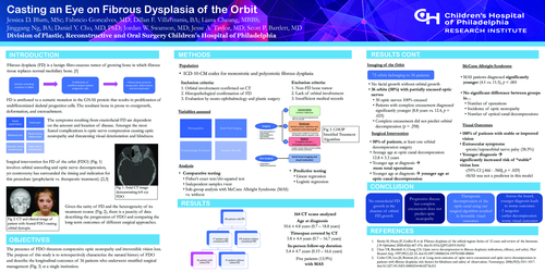
Surgical intervention for FD of the orbit (FDO) (Fig. 1) involves orbital unroofing and optic nerve decompression, yet controversy has surrounded the timing and indication for this procedure (prophylactic vs. therapeutic treatment). [2,3]
Given the rarity of FD and the heterogeneity of its treatment course (Fig. 2), there is a paucity of data describing the progression of FDO and comparing the long-term outcomes of different surgical approaches.
OBJECTIVES
The presence of FDO threatens compressive optic neuropathy and irreversible vision loss. The purpose of this study is to retrospectively characterize the natural history of FDO and describe the longitudinal outcomes of 36 patients who underwent stratified surgical management (Fig. 3) at a single institution.
METHODS
Population
Inclusion criteria:
1. Orbital involvement confirmed on CT
2. Histopathological confirmation of FD
3. Evaluation by neuro-ophthalmology and plastic surgery
Exclusion criteria:
1. Non-FD bone tumor
2. Lack of orbital involvement
3. Insufficient medical records
Analysis
RESULTS
164 CT scans analyzed
Age at diagnosis: 10.6 ± 4.8 years (0.7 – 18.8 years)
Timespan covered by CT: 3.8 ± 4.4 years (0.7 – 16.7 years)
In-person follow-up duration: 5.4 ± 4.7 years (0.15 – 16.6 years)
Five patients (13.9%) with MAS
Imaging of the Orbit
72 orbits belonging to 36 patients
McCune Albright Syndrome
Surgical Intervention
Visual Outcomes
-ptosis/supraorbital nerve palsy (38.5%)
-(95% CI [.466 - .960], p = .029)
-MAS was not a predictor in this model
CONCLUSION
1. No craniofacial FD growth in the absence of orbital FD growth
2. Progressive disease but complete encasement does not predict optic neuropathy
3. Therapeutic decompression of the optic canal using our surgical algorithm resulted in favorable visual outcomes
4. Across the board, younger diagnosis leads to worse outcomes
REFERENCES